Specimen Source
May be isolated from:
- Sputum
- Blood
- Vesicle fluid
- Stool
- Cerebrospinal fluid (CSF)
Note: These specimens present an exposure risk to the laboratorian.
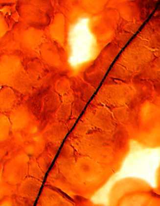
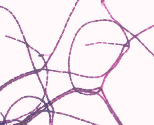
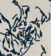
Large gram-positive spore-forming rods (1 to 1.5 by 3 to 5 microns) arranged in chains. Forms oval, central to subterminal spores that do not swell the bacterial cell. Spores are formed under atmospheric conditions. Spores are not present in clinical specimens.
Special Stain
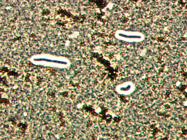
The India ink stain is used to stain capsules. Capsules appear as a well defined clear zone around the cell. Capsule formation may be evident in smears from clinical specimens, but not when the organisms are grown on most common laboratory culture media.
Colony Characteristics
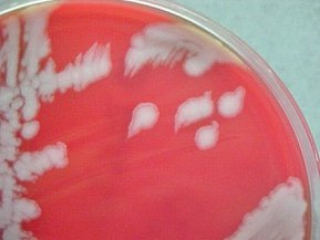
Sheep blood agar (SBA)
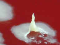
Colonies are nonhemolytic, flat or slightly convex with irregular edges and ground-glass appearance. There are often comma shaped projections from the colony edge producing a “Medusa-head” colony.
Colonies have a sticky consistency when manipulated with a loop.
No growth on MacConkey agar.
Motility
Bacillus anthracis is nonmotile. This is an unusual characteristic among Bacillus species.
Method 1: Wet Mount Slide
- Prepare a wet mount slide of a suspect colony from a 12 to 20 hour culture or from a fresh broth.
Method 2: Motility Test Medium
- Inoculate motility test medium. Incubate at 35 to 37 degrees C for 18 to 24 hours.
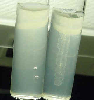
Motility test medium
Nonmotile Bacillus anthracis is on the right. Growth is a single line of growth that does not deviate from the original inoculum stab. Note the diffuse cloud of motility in the positive control on the left.
Motile Bacillus species with diffuse growth throughout the tube is on the right.
Key Characteristics
- Large, box-car shaped, gram-positive rod in short or long chains.
- Non-swelling, oval spores formed when grown on culture media. (Not usually seen in clinical specimens.)
- Encapsulated rods may be seen in clinical specimens.
- Ground-glass appearance of colonies.
- Nonhemolytic on sheep blood agar.
- Nonmotile.
- No growth on MacConkey.
- Catalase positive.
- Susceptible to penicillin.
- Lecithinase positive.
Identification pitfalls to avoid
Large, gram-positive bacilli that grow aerobically as nonhemolytic colonies on sheep blood agar should not be discarded as a contaminant, but should be worked up to rule out B. anthracis.
Look-a-likes
- B. megaterium (lecithinase negative, motile)
- B. thuringiensis (penicillin R, motile)
- Paenibacillus macerans (weak lecithinase, motile)
Safety
- Use biosafety level 2 practices for specimen processing.
- Use biosafety level 3 for activities with potential for aerosol or droplet production.
